A joint research team led by Dr. WANG Kai and Dr. DU Jiulin from the Institute of Neuroscience, Center for Excellence in Brain Science and Intelligence Technology of the Chinese Academy of Sciences reported a novel volumetric imaging method - confocal light field microscopy (Confocal LFM) - to image fast neural and vascular dynamics at high speed and deep in the brain.
The study was published in Nature Biotechnology on August 10.
Traditional tools developed for in-vivo imaging in the brain, such as confocal microscopy and two-photon scanning microscopy, are based on point scanning scheme and too slow to study rapid volumetric dynamics.
Light Field Microscopy (LFM) has attracted lots of attention due to its instantaneous volumetric imaging capability, which captures the information from the whole 3D volume all together on the image sensor in a single camera exposure.
Conventional LFM has been previously introduced to image brains. However, they suffer from two major problems: reconstruction artifacts and lack of optical sectioning capability, preventing them from wide applications.
In 2017, WANG's group developed a new type of LFM, called XLFM, to address the first problem. However, the second one remains unsolved.
Introducing optical sectioning capability in LFM is of key importance because it prevents out-of-focusing background from interfering with in-focusing signal and plays a vital role for thick tissue imaging.
Directly adopting the concept of confocal detection that helps confocal scanning microscope to gain 3D imaging capability seems to be straight forward. However, it turned out impossible due to the very distinct instantaneous volumetric imaging manner of LFM from conventional plane-by-plane imaging scheme.
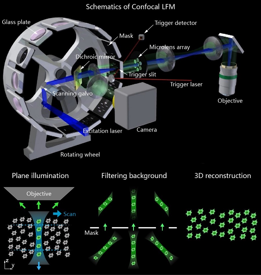
To meet this challenge, WANG's lab came up with a novel idea to generalize the concept of confocal detection and made it compatible with LFM without compromising its imaging speed, which was called Confocal LFM.

Fig. 1 Schematics of Confocal LFM. (Upper) Overview of the optical system. (Lower) Vertical light sheet scans in the sample plane and images formed by micro-lens array are spatially filtered by the mask to get in-focus volume after 3D reconstruction. (Image by CEBSIT)
Due to the elimination of interfering background, Confocal LFM significantly improved imaging resolution and calcium signal detection sensitivity in functional imaging of neuronal activities over the whole zebrafish brain.
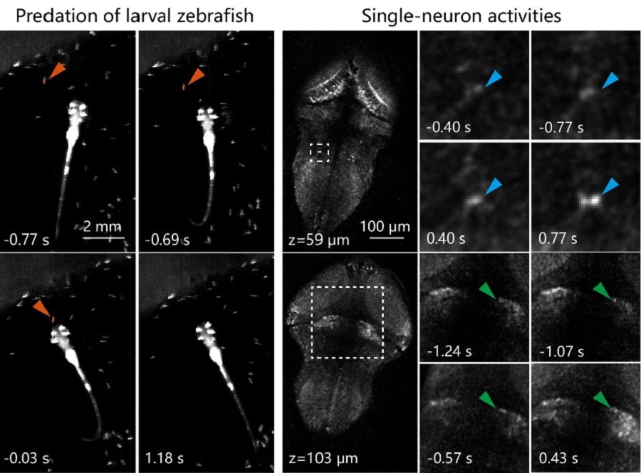
It was also integrated with a fast 3D tracking system to capture behavioral correlated neural activations in freely swimming larval zebrafish. For example, single-neuron activities during larval zebrafish’s prey capture could be reliably recorded.

Fig. 2 (Left) An example of larval zebrafish’s prey capture. Time was aligned to prey ingestion (0s). (Right) Zoomed-in views of single-neuron activities in several brain regions during prey capture in left. (Image by CEBSIT)
The researchers further applied Confocal LFM on imaging calcium transients and circulating blood cells in awake mouse brain. It’s highly parallelized and light efficient imaging manners enabled continuous recording for more than 100,000 volumes without significant photobleaching.
They also showed imaging of populations of circulate blood cells in a complicated 3D vascular network. The results showed that it was possible to track and quantify the flow dynamics of blood cells accurately and represented more than 100 times increase in throughput compared to conventional imaging techniques.
The flexibility, increased resolution and sensitivity of Confocal LFM demonstrated in this work made it promising for wide applications in capturing and studying extremely fast volumetric dynamics deep in the brain, or other types of thick tissues in general.

86-10-68597521 (day)
86-10-68597289 (night)

52 Sanlihe Rd., Xicheng District,
Beijing, China (100864)

