
Studies have shown that even a few hours of monocular deprivation can markedly improve the visual function of the deprived eye in adults. However, the underlying neural mechanisms of this ocular dominance plasticity remain unclear.
In a new study published in Communications Biology on November 11, a collaborative research team from the Institute of Biophysics of the Chinese Academy of Sciences and Wenzhou Medical University has revealed two opposing yet complementary mechanisms of ocular dominance plasticity operating in the subcortical and cortical regions of the adult visual system following short-term monocular deprivation.
Using 7T high-resolution functional MRI, the researchers examined changes in ocular dominance after three hours of monocular contrast deprivation in adult participants.
The results showed a significant enhancement in the deprived eye's contrast sensitivity and ocular dominance, accompanied by increased signals in the parvocellular layers of the lateral geniculate nucleus and the ventrolateral subdivision of the pulvinar.
At the same time, the non-deprived eye exhibited markedly improved 3D shape perception, along with stronger activation in stereopsis-related visual cortical areas. Interestingly, ocular dominance plasticity in the pulvinar and in the visual cortex was negatively correlated, exhibiting a reciprocal pattern of change.
These findings demonstrate that the adult brain harbors two opposing yet complementary mechanisms of ocular dominance plasticity. Together, these mechanisms allow the visual system to respond rapidly and adaptively to imbalances in binocular input.
This study provides important insights for developing intervention strategies for visual disorders such as amblyopia.
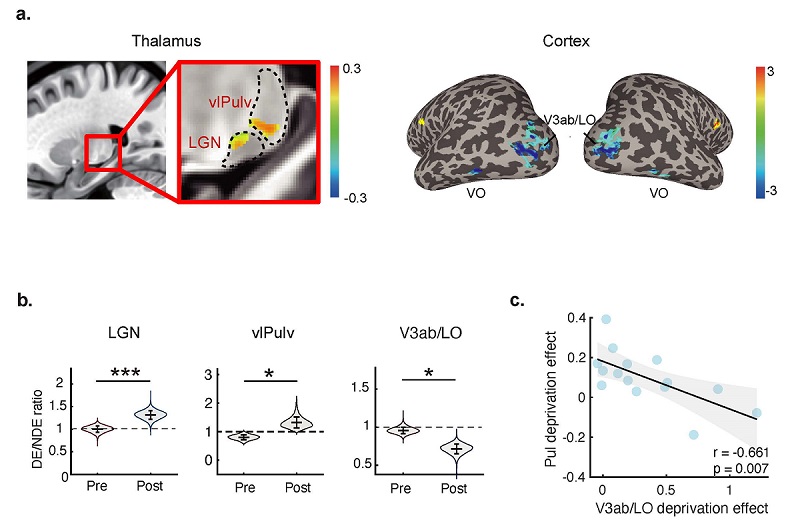
Two opposing but complementary mechanisms of ocular dominance plasticity in the visual thalamus and cortex (Image by ZHANG Peng's group)
Recently, Dr. BAO Min and his team from the Institute of Psychology of the Chinese Academy of Sciences and their collaborator Dr. HE Sheng from the Institute of Biophysics tested this idea.
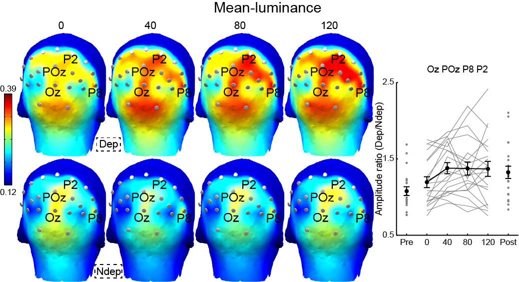
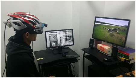
Patching one eye of an adult for a few hours, also termed short-term monocular deprivation (MD), has recently become an interesting research topic on visual plasticity. All the past work has measured the effects of MD using laboratory synthetic stimuli (e.g., gratings).

86-10-68597521 (day)
86-10-68597289 (night)

52 Sanlihe Rd., Xicheng District,
Beijing, China (100864)
Copyright © 2002 - Chinese Academy of Sciences


