Newsroom


Amblyopia, or "lazy eye," is a prevalent visual developmental disorder typically characterized by reduced vision in one eye. Previous studies have shown that signals from the amblyopic eye are weakened during transmission, the precise cortical circuit changes—particularly in feedforward, lateral, and feedback processing—have remained poorly understood.
In a collaborative study published in Imaging Neuroscience, researchers from the Institute of Biophysics of the Chinese Academy of Sciences and the Eye & ENT Hospital of Fudan University employed ultra-high-resolution 7 Tesla (7T) functional magnetic resonance imaging (fMRI) along with frequency-tagged electroencephalography (EEG) to reveal abnormal neural activity in the microcircuitry of the visual cortex in human patients with amblyopia.
The researchers found that in the primary visual cortex (V1) of amblyopic patients, visual signals from the amblyopic eye were significantly weakened as early as the input layer-an intracortical sublayer primarily responsible for receiving thalamic input-and that this weakened signal was subsequently transmitted forward to downstream visual areas, suggesting that the abnormalities in amblyopia originate from a very early stage of visual information input.
Furthermore, an imbalance in the lateral inhibition mechanisms between the two eyes led to a loss of signals in the superficial layers of V1. The dominant (non-amblyopic) eye exerted strong inhibitory effects on the amblyopic eye's signal transmission, while the amblyopic eye showed significantly reduced inhibition of the dominant eye.
Frequency-tagged EEG data further indicated that this imbalance in interocular inhibition was accompanied by a marked decline in the brain's ability to integrate visual information from both eyes. In addition to reduced signal amplitude, visual signals from the amblyopic eye also showed noticeably slower transmission, indicating an overall decline in visual processing efficiency.
This study is the first to characterize amblyopia-related dysfunction in the human visual cortex at sub-millimeter spatial resolution and millisecond temporal resolution. It demonstrates how disrupted visual experiences during critical periods of development can alter the architecture and function of cortical microcircuits.
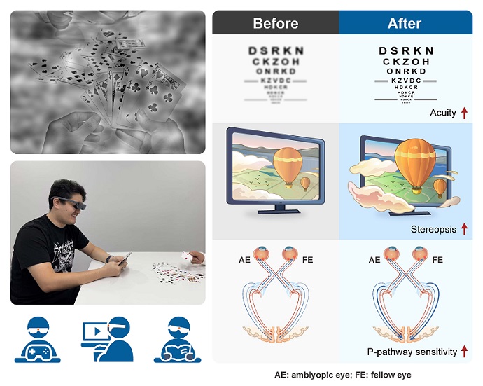
The results offer significant insights into the neural mechanisms underlying amblyopia and provide a new theoretical foundation for treatment. "Visual training aimed at enhancing the function of input-layer neurons, improving binocular information integration, and correcting imbalanced inhibition may represent new directions for future amblyopia therapy," said Prof. ZHANG Peng, first author of the study.
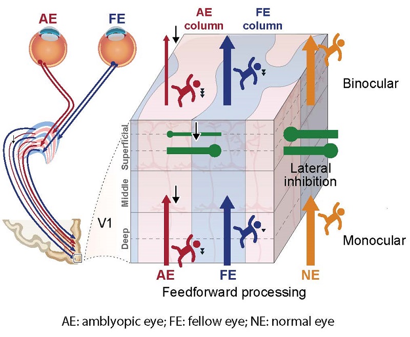
Visual signals from the amblyopic eye are already impaired upon entering the cortex. Binocular inhibition is imbalanced, integration is weakened, and signal transmission is delayed. (Image by ZHANG Peng's group)