
Adult stem cells have long been considered as a very promising candidate cell source for regenerative medicine. They are non-tumorigenic, relatively safe, and less controversial without any embryo manipulation. Indeed, as an example of adult stem cell therapies, hematopoietic stem cell (HSC) transplantation has been practiced in clinic for decades. The ideal strategy of adult cell therapy would start with taking a small biopsy from the patient/donor. The stem cells will then be isolated and expanded in vitro. The in vitro expansion will yield sufficient amount of stem cells for transplantation.
However, the utilization of this strategy has been hampered by the lack of efficient in vitro system to expand functional adult stem cells. The majority of the adult stem cells, including HSCs and muscle stem cells (MuSCs), are unable to be expanded in vitro. In the case of muscle stem cells, these cells can barely be cultured and expanded in vitro. Ex vivo cultured MuSCs differentiate to myoblast progenitor cells in a few days and lose most of their abilities to regenerate new muscles in vivo. Due to this reason, the initial clinical trials of myoblast progenitor transplantation failed. Establishment of an in vitro system to culture and expand functional MuSCs will break this bottleneck and capitalize the advantage of adult stem cell based therapies.
Recently, Prof. HU Ping and Prof. WANG Hongyan’s research groups from Institute of Biochemistry and Cell Biology, Shanghai Institutes for Biological Sciences solved the in vitro MuSCs expansion problem using an elegant method. They started their investigation by characterizing the endogenous microenvironment of MuSCs proliferation and expansion. They identified T cells as a critical MuSCs microenvironment component. MuSCs proliferation was greatly promoted by co-culturing with T cells.
By mimicking the endogenous microenvironment in vitro, functional MuSCs can be expanded for over 20 passages in vitro. In a collaborative investigation, HU and WANG’s groups further nailed the minimum effective components of cytokines secreted by T cells down to four cytokines. The four cytokine cocktail could facilitate MuSCs proliferation and expansion in vitro. MuSCs cultured in medium supplemented with the four cytokines can maintain their stemness for over 20 passages. The starting number of MuSCs was increased for over 1018 fold in this in vitro expansion system. The in vitro expanded MuSCs express all the MuSCs markers. They can repair muscle injury in vivo efficiently after cell transplantation. The transplanted MuSCs are capable of homing to the right niche and replenishing the endogenous MuSCs pool. After a single transplantation, the exogenous MuSCs are able to repair multiple rounds of muscle injuries.
These results showed that T cell mediated acute inflammation provides an indispensable microenvironment for MuSCs expansion. It may provide new guidance for the usage of anti-inflammation drugs after trauma. Furthermore, the establishment of the in vitro system to expand functional MuSCs for long term can provide sufficient amount of MuSCs for potential regenerative medicine. It marks a big step towards treating muscle atrophy and injuries by MuSCs based therapy. It also provides new strategies to expand other types of adult stem cells in vitro. The study was published in Cell Research as cover story.
This project received supports of the Chinese Academy of Sciences, the Ministry of Science and Technology of China, the Science and Technology Commission of Shanghai Municipality, and the National Natural Science Foundation of China.
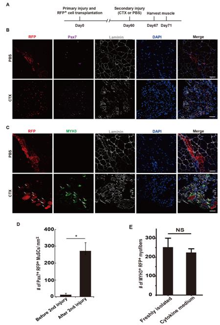
MuSCs treated with cytokine cocktail support the secondary injury repair after a single transplantation. (A) Strategy to examine the abilities of MuSCs to repair the secondary injury with a single transplantation. Primary injury and MuSC transplantation were performed at TA muscles in hindlimb using wild type recipient mice. The secondary injury was induced 60 days later. Muscle tissues were harvested at 7 and 11 days post the secondary injury to analyze the proliferative capabilities of RFP+ cells and their abilities to repair injury. (B) Pax7 staining of sections from TA muscle 7 days after the secondary injury. RFP+ MuSCs expanded in cytokine medium were transplanted into injured wild type non-fluorescent recipient TA muscle as described in Figure 5c. The secondary injury was induced 60 days later. The proliferation ability of RFP+ MuSCs upon the secondary injury stimulation was analyzed by immunofluorescent staining of Pax7 and observed by confocal microscopy. Red indicated RFP; Purple indicated Pax7 immunofluorescent staining; Grey indicated laminin immunofluorescent staining; Blue indicated DAPI staining of nuclei; Merge indicated merged images of Pax7, Laminin, DAPI staining and RFP. Scale bars, 20μm. (C) MYH3 staining of sections from TA muscle 11 days after the secondary injury. The newly formed myofibers were monitored by MYH3 immunofluorescent staining. Newly formed myofibers derived from transplanted MuSCs should be both MYH3+ and RFP+. Red indicated RFP; Green indicated MYH3 immunofluorescent staining; Grey indicated laminin staining; Blue indicated DAPI staining of nuclei; Merge indicated merged images of MYH3, laminin, DAPI staining and RFP. Scale bars, 20 μm. (D) Statistical analysis of the number of RFP+Pax7+ MuSCs 7 days after the second injury. Error bars were based on 4 independent experiments. * indicated statistically significant. P<0.01. (E) Statistical analysis of the reparation efficiency after the secondary injury. Error bars were based on 4 independent experiments. NS indicated no statistically significant difference. (Image by Prof. HU Ping`s group)
CONTACT:
HU Ping, Principal Investigator
Institute of Biochemistry and Cell Biology, Shanghai Institutes for Biological Sciences, Chinese Academy of Sciences
Shanghai 200031, China
Phone: 86-21-54921254
E-mail: hup@sibcb.ac.cn

86-10-68597521 (day)
86-10-68597289 (night)

86-10-68511095 (day)
86-10-68512458 (night)

cas_en@cas.cn

52 Sanlihe Rd., Xicheng District,
Beijing, China (100864)
Copyright © 2002 - Chinese Academy of Sciences


