Newsroom


In human, the ovary is one of the organs exhibiting early-onset aging-associated dysfunction. As a critical female reproductive organ, the ovary consists of numerous heterogeneous cell types including follicles at different developmental stages. To date, however, it is difficult to accurately reveal the molecular mechanisms of human ovarian aging, and our understanding of human ovarian aging and the development of interventions against human ovarian aging and associated diseases are hindered.
Recently, scientists from the Institute of Zoology of the Chinese Academy of Sciences, Peking University, and Salk Institute for Biological Studies have worked jointly and established the first single-cell transcriptomic atlas of primate ovarian aging.
Using the single-cell transcriptome sequencing technology, they drew a comprehensive aging roadmap of cynomolgus monkey ovaries at the single-cell resolution. Through the bioinformatic analyses of the single-cell transcriptomic data of ovarian tissues from monkeys and the experimental validation in human ovarian cells, they identified the disturbance of antioxidant signaling as a main feature of primate ovarian aging.
This study entitled "Single-cell transcriptomic atlas of primate ovarian aging" is published online in Cell on January 30, 2020.
The researchers first obtained ovarian tissues from young and old cynomolgus monkeys. Histopathological analysis revealed increased atretic follicles, decreased healthy follicles, and increased fibrosis in aged ovaries, suggesting that the primate ovaries underwent aging-associated structural and functional degenerative changes. By the modified single-cell tagged reverse transcription (STRT) single-cell transcriptome sequencing, the single-cell transcriptomic landscape of ovaries from young and aged non-human primates (NHPs) was depicted and seven ovarian cell types (including oocyte and six types of ovarian somatic cells) with distinct gene expression signatures were identified.
In-depth dissection of gene expression dynamics of oocytes revealed four subtypes at sequential and stepwise developmental stages. Further study uncovered that aging led to the imbalance of cell type-specific redox regulatory networks in the ovary. Antioxidant genes including GPX1 and GSR were specifically downregulated in aged early-stage oocytes, suggesting that early oocytes were susceptible to oxidative stress.
In addition, pro-apoptotic genes were upregulated while oxidoreductase-related genes were downregulated in aged granulosa cells (GCs) that interact with oocyte and regulate oocyte development during folliculogenesis. Moreover, genes involved in redox regulation, such as IDH1 and PRDX4, were identified as new aging biomarkers for GCs.
The researchers then analyzed human GCs from the follicular fluid of healthy women undergoing assisted reproduction technology, revealing age-dependent increases in reactive oxygen species and apoptosis, as well as decreases in the aforementioned aging hallmarks in human GCs.
In addition, a causal link between the changes in redox gene expression and oxidative damage in human GCs was identified, indicative of similar cellular and molecular events during human ovarian aging as observed in cynomolgus monkeys.
Taken together, the researchers established a high-resolution single-cell transcriptomic atlas of non-human primate ovarian aging and identified cell type-specific dysregulation of redox homeostasis as a major feature of primate ovarian aging.
This work deepens the understanding of aging phenotypes of ovarian tissues, reveals the susceptibility and susceptible molecules of different ovarian cell types to aging, provides new diagnostic biomarkers and potential therapeutic targets for age-related human ovarian disorders, and opens new avenues for developing novel tools for aged oocyte rejuvenation for assisted reproductive therapies and female fertility preservation.
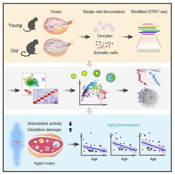
Figure. Single-Cell Transcriptomic Analysis of Primate Ovarian Aging (Image by LIU Guanghui)