
Newsroom


Losing one immune gene turns male and female bodies into two different battlefields. Recently, a research group led by Associate Prof. WU Nan and Prof. XIA Xiaoqin from the Institute of Hydrobiology (IHB) of the Chinese Academy of Sciences (CAS) has revealed how foxp3a controls sex-dimorphic immune-metabolic regulation across the gut-liver-gonad axis in zebrafish. The findings were published in Fish and Shellfish Immunology.
Sex differences in intestinal mucosal immunity are a well-recognized immunological phenomenon, but the underlying mechanisms and their links to other organs remain poorly understood. The gene foxp3a, a master regulator of regulatory T cells (Tregs), has been linked to intestinal inflammation and testicular gametogenesis in zebrafish, positioning it as a key candidate for studying sex-specific immune regulation across organ systems.
By examining foxp3a loss-of-function mutants across three developmental timepoints, the researchers found sex-dependent alterations in intestinal mucosal barrier integrity, goblet cell patterns, and immune cell composition. In males, innate immune and inflammatory pathways were persistently activated; in females, compensatory metabolic and microbiota-based responses protected the mucosa and limited inflammation.
Single-cell transcriptomic sequencing of intestinal mucosal cells revealed Treg depletion and Th1/Th17 expansion in both sexes. In female dendritic cells only, tight junction pathways were upregulated, whereas MAPK signaling, apoptosis, and C-type lectin receptor signaling were simultaneously downregulated. This suppressed antigen presentation and correlated with reduced CD8+ T and tissue-resident memory T cells. Cell-cell communication analysis showed stronger goblet cell–dendritic cell interactions in males, amplifying inflammation, while female epithelial–innate immune crosstalk actively suppressed inflammatory escalation.
In the liver, female mutants displayed autophagy and endoplasmic reticulum stress, whereas males exhibited hepatocyte swelling and lipid metabolic dysregulation.
In the gonads, ovaries displayed mTOR suppression consistent with delayed oogenesis, whereas testes showed stronger immune activation and tissue damage, echoing the team's earlier finding that foxp3a deficiency causes male infertility in zebrafish.
"The loss of a single immune regulatory gene triggers fundamentally different responses in males and females, reshaping the gut, liver, gonads, and microbiome in distinct ways," WU said. "This points to foxp3a as a sex-biased coordinator of whole-body homeostasis."
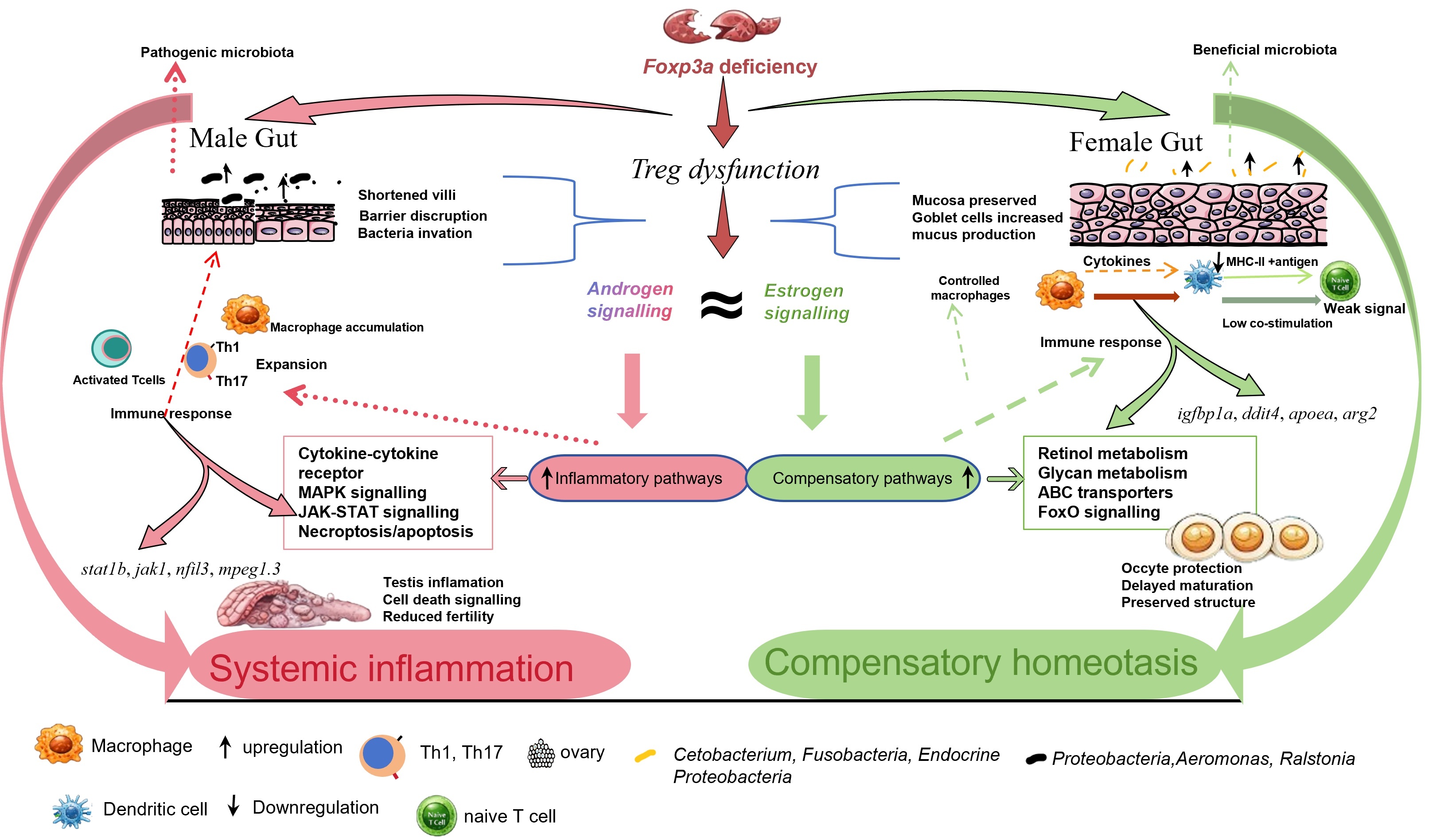
Sex dimorphism in foxp3a-mediated immune regulation and associated metabolic processes along the gut-liver-gonad axis. (Image by IHB)
Cross-organ co-expression analysis revealed sex-specific inter-organ gene coordination.
Female modules contained mucosal repair gene igfbp1a and stress regulator ddit4, while liver-ovary co-expressed genes linked complement-mediated immune dysregulation to delayed oogenesis. Male modules were enriched in inflammation genes including jak1, stat1b, and nfil3, and gut-testis co-expressed genes connected mucosal repair to spermatogenesis, suggesting an intrinsic link between foxp3a-controlled intestinal barrier integrity and gonadal development.
At the host-microbiota level, foxp3a deficiency altered gut microbial composition in a sex- and age-dependent manner. Microbiota dysbiosis was linked to gonadal function: in females, the ovarian development gene rpl10 correlated negatively with Proteobacteria; in males, Finegoldia correlated negatively with reproductive genes (tdrd7a, dazl, piwil1) but positively with MHC class I components, revealing a microbial link between gut dysbiosis and gonadal immune dysfunction.
The researchers suggest that foxp3a contributes to the sex-biased coordination of immune, metabolic, and reproductive processes across organs, positioning it as a broad integrator of whole-body homeostasis beyond its classical role as a T cell transcription factor. The sex-dependent mechanisms identified here may provide broader insight into why males and females differ in susceptibility to immune-metabolic disorders.