2024


Recent years have seen an increasing demand for cross-scale high-throughput imaging, however, conventional microscope objectives cannot simultaneously meet the requirements for large field and high-resolution imaging.
Mesoscale microscope objectives, with complex optical structures and excellent aberration optimization, can achieve both high numerical aperture and ultra-large field of view (FOV), and significantly enhance the imaging throughput of optical microscopes.
Recently, a group led by SHI Guohua from Suzhou Institute of Biomedical Engineering and Technology (SIBET) designed a flat-field apochromatic objective lens structure under a mesoscale field and developed a mesoscale microscope objective with the largest reported FOV and the widest working band at sub-micron resolution.
Objective lens is the core component of optical microscope, determining two critical parameters of microscopic imaging: resolution and imaging field of view (FOV). The resolution and FOV of the objective lens are interdependent. FOV of existing mesoscale objectives is concentrated between 3mm and 6mm diameter.
This objective lens features an 8mm FOV diameter, a 0.5NA numerical aperture, and an imaging band extending from 400-1000nm. Utilizing this objective lens for imaging mouse brain and kidney slices, researchers obtained ultra-high-throughput images with a single frame of 1.35 billion pixels (Figure 2a).
Current mesoscale objectives are limited to a single imaging wavelength, capable of only visible or near-infrared single-band imaging, and cannot meet the requirements for diverse fluorescence imaging.
The researchers constructed a related imaging system (Figure 1) and achieved single and two-photon mesoscale imaging, for the first time, using the same objective lens (Figure 2b).
A quantitative comparison with a commercial 20x 0.5 NA objective lens indicated that this mesoscale objective had similar imaging quality to the commercial lens but provided an FOV over 40 times larger.
Experimental results demonstrate that this objective lens has significant potential for large-scale sample high-resolution multi-wavelength imaging, such as brain mapping, cross-brain region single and two-photon imaging, and high-resolution imaging of organoids.
Results of the study entitled “Large-field objective lens for multi-wavelength microscopy atmesoscale and submicron resolution” were published in the recent issue of Opto-Electronic Advances.
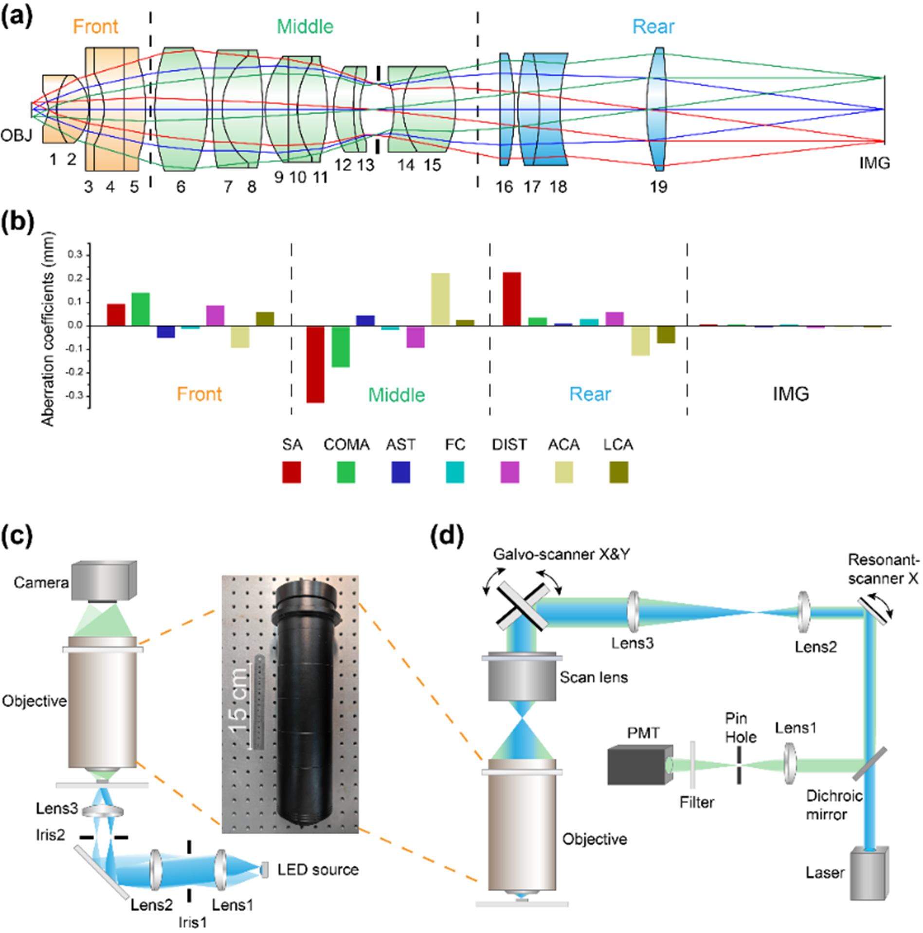
Figure 1. Mesoscopic objective lens structure and imaging system configuration. (Image by SIBET)
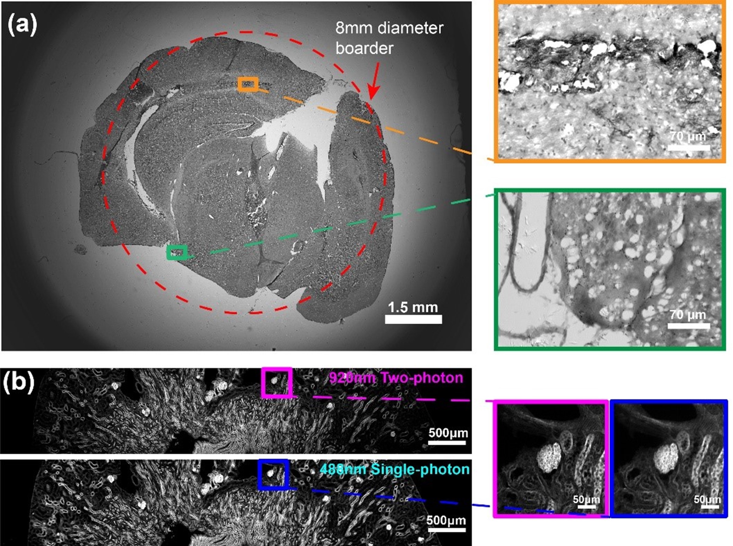
Figure 2. Imaging results of biological samples. (Image by SIBET)