
Magnetic resonance imaging (MRI) is an important tool for defining fetal anatomy and pathology non-invasively, especially for the diagnosis of neurodevelopmental disorders and cardiovascular diseases.
Due to a lack of fetal imaging coils, the standard commercial abdominal coil is often used for fetal imaging. However, its performance is limited by insufficient coverage, element number and signal-to-noise ratio (SNR).
A research team led by Prof. ZHENG Hairong at the Shenzhen Institutes of Advanced Technology (SIAT) of the Chinese Academy of Sciences developed a dedicated 36-channel coil array for fetal imaging at 3 Tesla, with high density coil elements, wide coverage and flexibility to conform to the varied anatomy of pregnant patients.
Compared to a commercial abdominal coil array, the proposed 36-channel fetal array provides not only SNR improvements up to 40%, but also an augmented imaging speed. Clear fetal brain (twice as fast) and heart movie (4 times faster) imaging were successfully performed with the dedicated 36-channel coil array.
The research entitled "A Dedicated 36-channel Receive Array for Fetal MRI at 3 T" was published in IEEE Transactions on Medical Imaging.
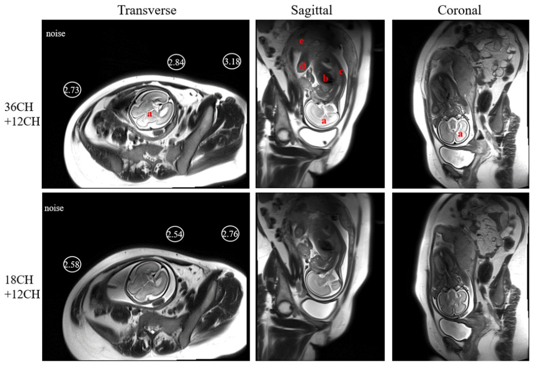
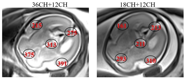
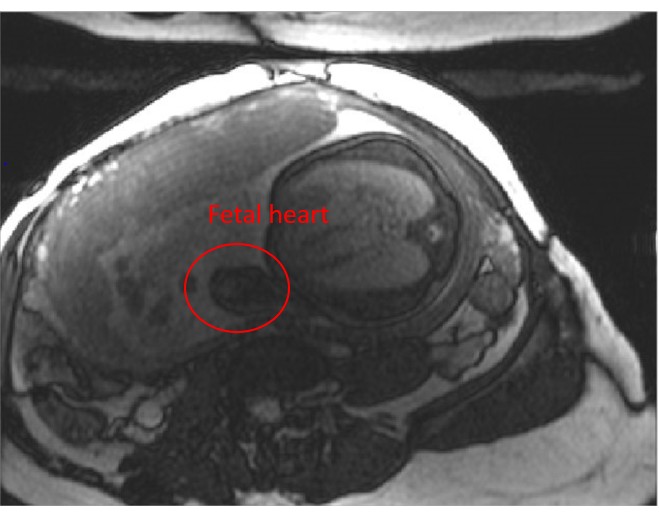
Figure 2. Fetal heart image in the transverse planes acquired by using the proposed 36-channel coil combined with a 12-channel spine coil. (Image by ZHENG Hairong)

86-10-68597521 (day)
86-10-68597289 (night)

86-10-68511095 (day)
86-10-68512458 (night)

cas_en@cas.cn

52 Sanlihe Rd., Xicheng District,
Beijing, China (100864)
Copyright © 2002 - Chinese Academy of Sciences


