Chinese researchers said Wednesday they have found direct evidence that Zika infection causes microcephaly, a birth defect marked by an abnormally small head, in mouse experiments.
Mouse fetuses injected with the Zika virus and carried to term within their pregnant mothers display the characteristic features of microcephaly, they reported in the U.S. journal
Cell Stem Cell.
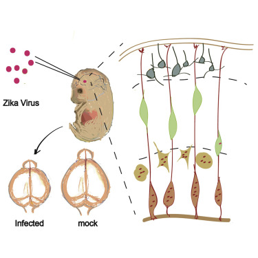
As expected, the virus infected the neural progenitor cells, and infected brains revealed expression of genes related to viral entry, altered immune response, and cell death.
The research was a collaborative effort between XU Zhiheng at the Institute of Genetics and Developmental Biology of the Chinese Academy of Sciences and QIN Cheng-Feng at the Beijing Institute of Microbiology and Epidemiology.
"The most surprising part of this study is that it was mostly neural progenitor cells that got infected in the beginning and mostly neurons that became infected at a later stage -- five days after injection when the presence of Zika virus increases several hundred folds," said XU.
"However, almost all cell death was found in neurons other than neural progenitor cells. This indicates that neurons, but not neural progenitor cells, are prone to induced cell death by the Zika virus."
In the study, the Zika virus was injected directly into fetal mouse brains, and the researchers found that the embryos won't survive until it was given on the 13.5th day of pregnancy.
Total gestational period in the mouse is about 20 days long, so the time is equivalent of the second trimester in humans, when the fetus's neural progenitor cells are intensively expanding and generating new neurons at the same time, they said.
One problem they encountered was all the infected pups who survived to birth were eaten by their mothers.
"This indicates that the pups were too sick," said XU. "We have to use lower dose of Zika infection in the future to determine whether the pups will survive longer in order to determinate the long-term consequences of Zika infection on brain development."
To overcome this problem that prevented further observation, they dissected the pregnant mice to inspect the pups on the 18.5th day of pregnancy and found their heads were about a third smaller than those of normal pups.
"Therefore, this is the direct evidence that Zika infection causes microcephaly," XU said, but cautioning: "Mouse is not human, and we need be careful when translate our findings into human disease."
Next, the researchers planned to work with other institutes to use their mouse model to test different drugs and a vaccine against Zika.
In a second study, researchers at University of California San Diego, with colleagues in Brazil and Senegal, also said they found the first "direct experimental proof" that the Brazilian strain of Zika virus can cause severe birth defects.
The findings, published Wednesday in the British journal Nature, were based on research in mouse models, human stem cells and cerebral organoids, or miniature brains grown in vitro.
A third study using wide-type mice, which was published Wednesday in the U.S. journal Cell, said that Zika migrated from the pregnant mouse's bloodstream into the placenta, where it multiplied, then spread into the fetal circulation and infected the brains of the developing pups.
"Although wild-type adult mice are not the good host for Zika virus, their studies provide important evidence that immune response in the mother would also affect the development of fetus that may cause microcephaly," XU said of the two studies. (Xinhua)
Zika virus disrupts neural progenitor development and leads to microcephaly in mice. (Image by LI Cui, et al.)