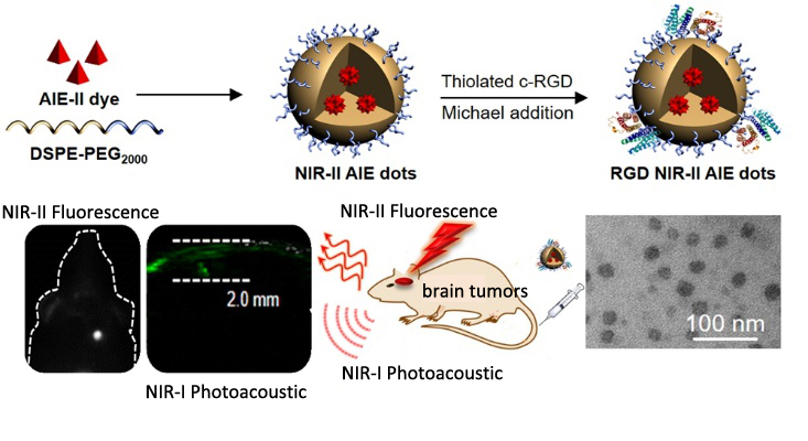
Nowadays, tumors inside the complex central nervous system remain one of the most challenging cancers to diagnose.
Different from conventional brain-imaging techniques, nearinfrared (NIR) fluorescence imaging demonstrates particular merits including being nonhazardous, offering fast feedback, and having higher sensitivity.
A research team led by Prof. ZHENG Hairong from the Shenzhen Institutes of Advanced Technology (SIAT) of the Chinese Academy of Sciences, in collaboration with Prof. LIU Bin from the University of Singapore, reported the first NIR‐II fluorescent molecule with aggregation-induced-emission (AIE) characteristics for dual fluorescence and photoacoustic imaging. Their findings were published in Advanced Materials.
Fluorescence imaging in the second NIR window (NIR-II), compared with the first NIR window (NIR-I), exhibits salient advantages of deeper penetration and higher spatiotemporal resolution, owing to further reduced photon scattering, absorption, and tissue autofluorescence in biological tissues.
Scientists designed a new donor-acceptor (D-A)-tailored NIR-II emissive AIE molecule, and formulated dots showed a high NIR‐II fluorescence quantum yield up to 6.2%, owing to the intrinsic aggregation‐induced emission nature of the designed molecule.
The AIE dots have been successfully used for dual NIR‐II fluorescence and NIR‐I photoacoustic imaging for precise noninvasive brain‐tumor diagnosis. Based on the same dots, the experiments revealed that NIR‐II fluorescence imaging showed a high resolution.
Meanwhile, NIR‐I PA imaging intrinsically exhibited higher penetration depth than that of NIR‐II fluorescence imaging, which allowed clear delineation of tumor depth in the brain.
The synergetic bimodal imaging with targeting c‐RGD‐decorated bright AIE nanoparticles showed precise brain‐tumor diagnosis with good specificity and high sensitivity, which yielded a high S/B of 4.4 and accurately assessed the depth of tumor location inside brain tissue.

The study demonstrates the promise of NIR-II AIE molecules and their dots in dual NIR-II fluorescence and NIR-I photoacoustic imaging for precise brain cancer diagnostics.
The research was supported by National Basic Research Program of China (973 Program), National Natural Science Foundation of China, Guangdong Province Magnetic Resonance Imaging and Multimode Systems Key Laboratory.

86-10-68597521 (day)
86-10-68597289 (night)

86-10-68511095 (day)
86-10-68512458 (night)

cas_en@cas.cn

52 Sanlihe Rd., Xicheng District,
Beijing, China (100864)

